The STTARR's histopathology core now provides 100+ optimized antibodies for immunohistochemistry/immunofluorescence. The histopathology core now specializes in the special MOVAT stain (highlights the various constituents of connective tissue, especially cardiovascular tissue, by five colors in a single stained slide). The new development of an in-situ hybridization protocol combined with immunofluorescence or human X-Y chromosome assays is now a histopathology service offered at STTARR.
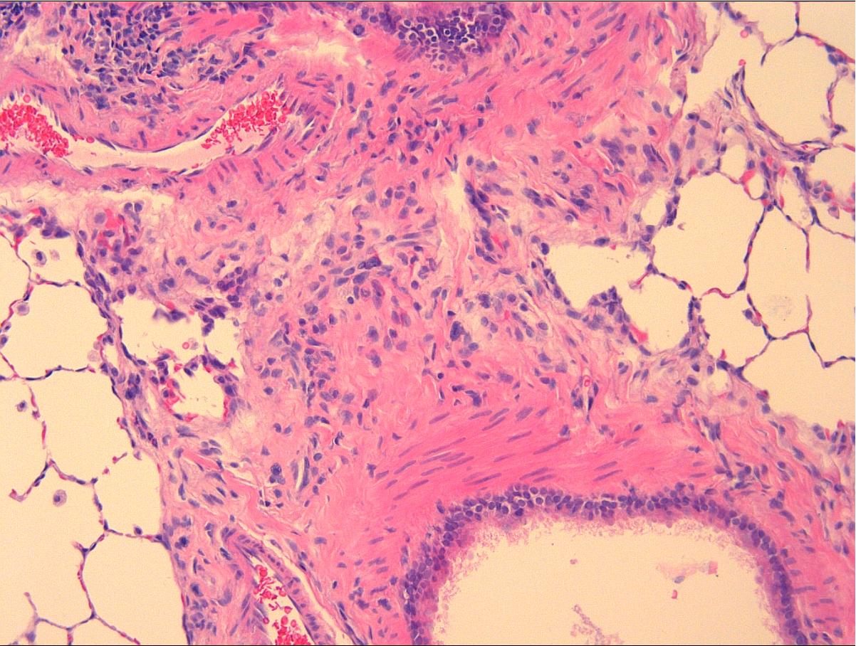
Hematoxylin and eosin staining on a lung tissue
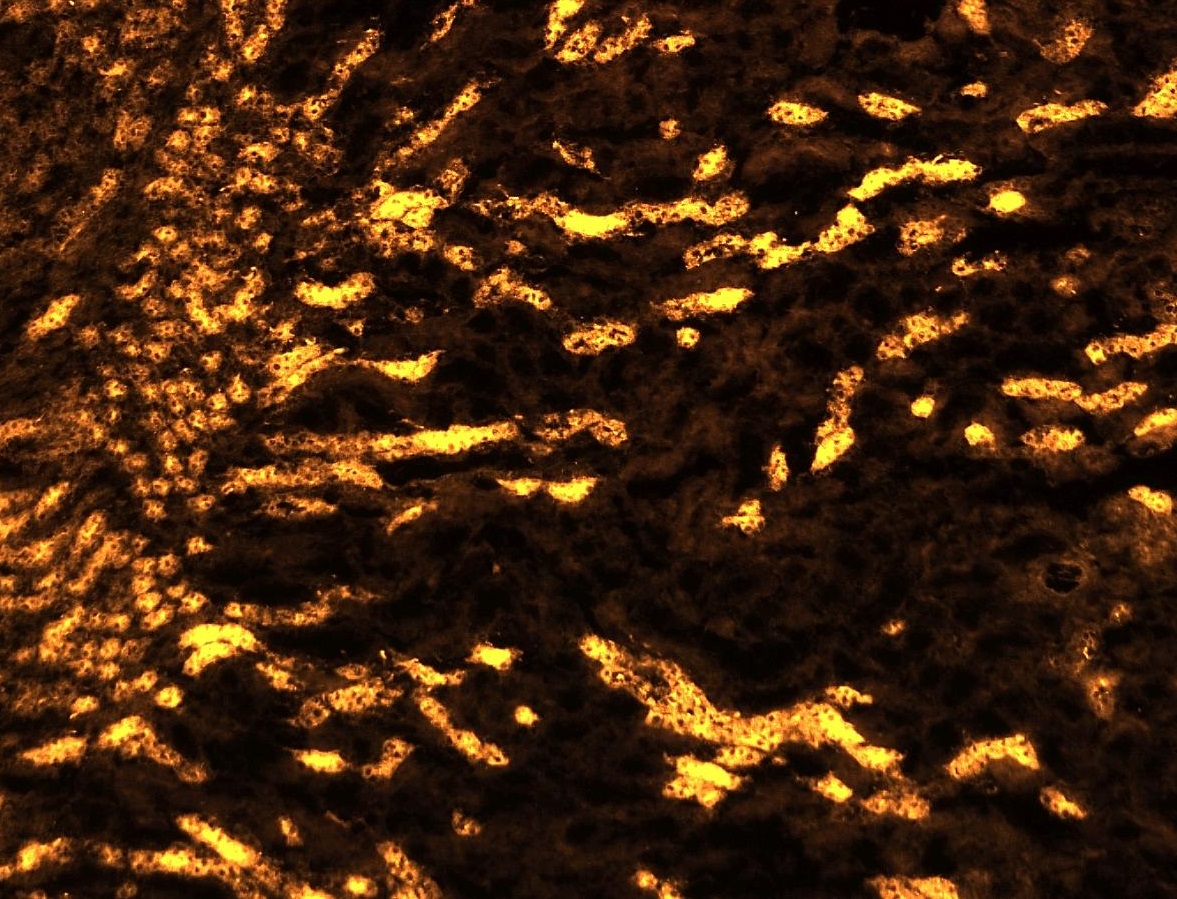
Kidney tubules marker stained by IF
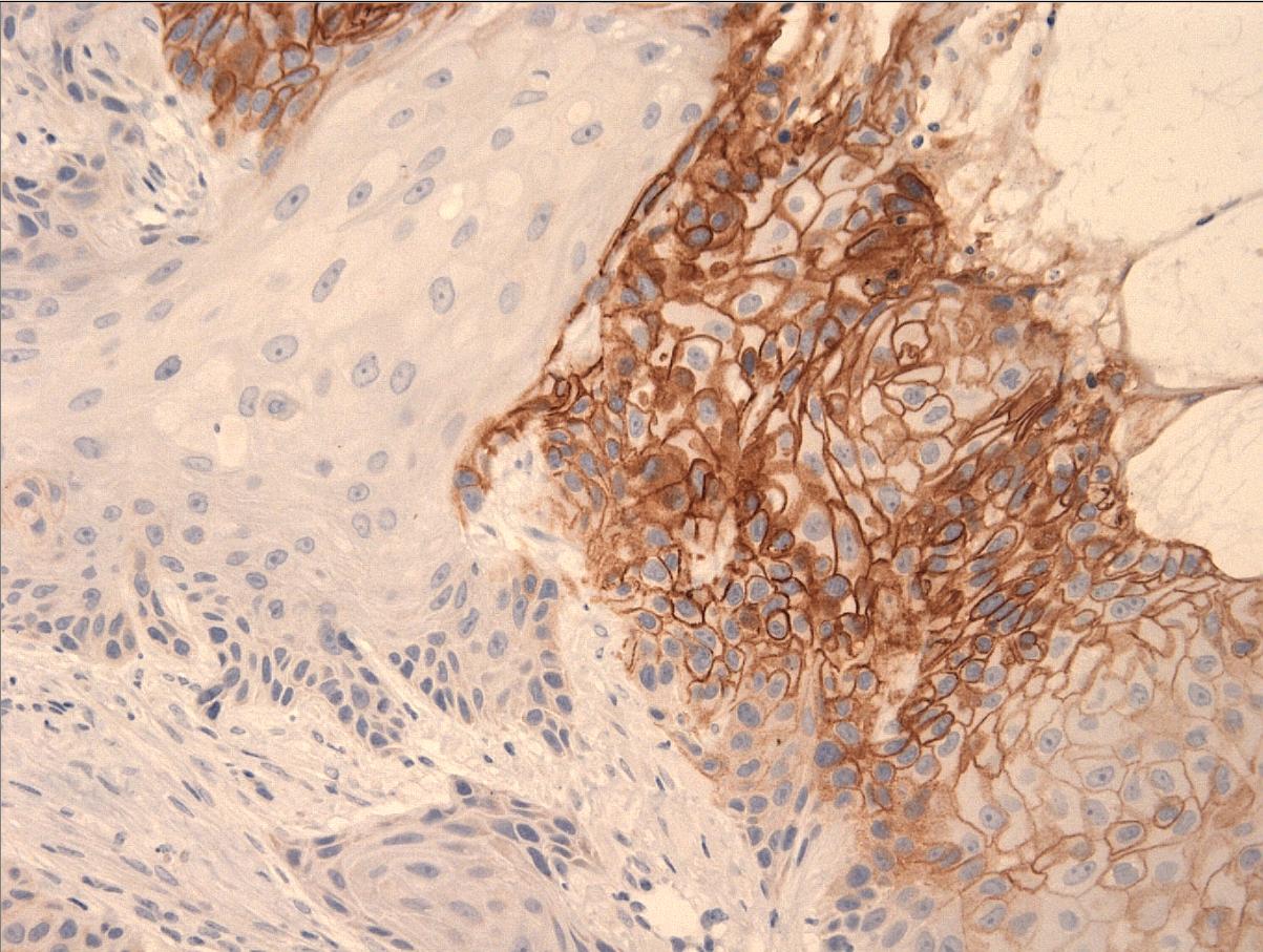
CA9 IHC for hypoxia screening
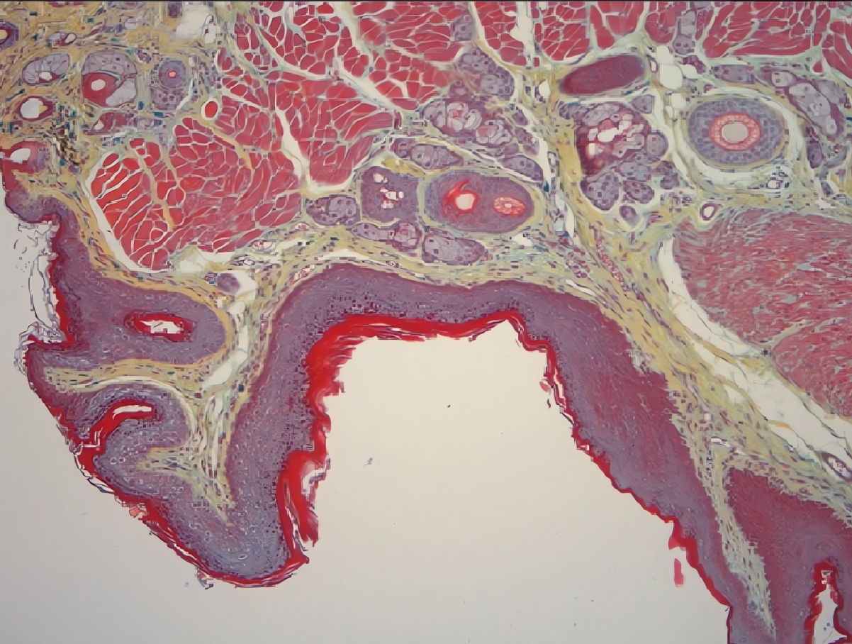
Movat pentachrome
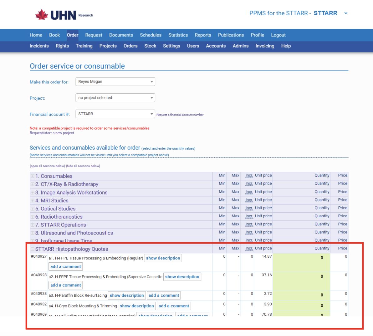
Assess sample fixation, trimming harvest tissue to the plane of interest.
Regular cassette size – 30mm x 35mm.
Supersize cassette size – 65mm x 50mm.
Melt down paraffin block and re-orient the tissue into the desired cutting plane.
Sectioning OCT frozen tissue blocks prepared by users.
User provide fresh harvested tissue to STTARR Histopathology for snap freezing in OCT.
Special snap freezing technique for muscle tissue prone to freeze artifact.
Sectioning paraffin and frozen section at routine 4um thickness or in special thickness requested by user.
Regular slides – 25mm x 75mm.
Wholemount large slides – 50mm x 75mm.
Paraffin or frozen (per slide) consecutive sections from a ribbon of section mounted on slides labelled with numbers.
Look for specific microscopic structures in paraffin/frozen blocks and take sections eg. Mouse aortic arch, aortic root, mouse embryo thyroid glands.
Optimization of commercial and/or in house produced antibodies.
Check out our Histopathology Optimized Antibody List for a list of antibodies currently available and optimized at STTARR Histopathology. Please note that the list is continuously updated. If the antibody you are interested in is not on the list, please contact us.
We also offer validated antibodies for immunohistochemistry staining on porcine tissue.
Multiplex of up to 3 target proteins in cells/tissues using fluorescent tagged antibodies.
Detection of DNA fragmentation in last phase of apoptosis (cell death).
Utilizing ACDBio System probes and detection systems to detect target mRNA in paraffin embedded sections.
Masson trichrome, Elastic trichrome, Picrosirius
red, Periodic acid Schiff, Cresyl echt violet, Luxol
fast blue, Gram stain, Von Kossa calcium, Oil O red,
Gordon & Sweets Silver Reticular fiberstain,
Safranin O, MOVAT, Prussian blue, Fontana Masson and
fast green, TRAP (Tartrate-Resistant Acid
Phosphatase).
STTARR Histopathology has other histochemistry stain
protocols that are available upon request, please
inquire if the stain that you are interested is not
on the list.
Principle and routine stain used in histopathology performed on paraffin or frozen sections.
Sectioning thick sections (3–50 microns) for autoradiography, 2 sets of slides each sample. Protocol on sample preparation for autoradiography available upon request.
Centrifuge cell pellet and double embed into agar gel and paraffin.
Formic acid for rapid decalcification or EDTA for tissue to be used for IHC.
Harvest tissue for toxicology study or specific organ of interest.
Combining multiple donor tissue paraffin blocks into one for high throughput IHC staining.
The STTARR's histopathology core now provides 100+ optimized antibodies for immunohistochemistry/immunofluorescence. The histopathology core now specializes in the special MOVAT stain (highlights the various constituents of connective tissue, especially cardiovascular tissue, by five colors in a single stained slide). The new development of an in-situ hybridization protocol combined with immunofluorescence or human X-Y chromosome assays is now a histopathology service offered at STTARR.
Hematoxylin and eosin staining on a lung tissue
Kidney tubules marker stained by IF
CA9 IHC for hypoxia screening
Movat pentachrome
Assess sample fixation, trimming harvest tissue to the plane of interest.
Regular cassette size – 30mm x 35mm.
Supersize cassette size – 65mm x 50mm.
Melt down paraffin block and re-orient the tissue into the desired cutting plane.
Sectioning OCT frozen tissue blocks prepared by users.
User provide fresh harvested tissue to STTARR Histopathology for snap freezing in OCT.
Special snap freezing technique for muscle tissue prone to freeze artifact.
Sectioning paraffin and frozen section at routine 4um thickness or in special thickness requested by user.
Regular slides – 25mm x 75mm.
Wholemount large slides – 50mm x 75mm.
Paraffin or frozen (per slide) consecutive sections from a ribbon of section mounted on slides labelled with numbers.
Look for specific microscopic structures in paraffin/frozen blocks and take sections eg. Mouse aortic arch, aortic root, mouse embryo thyroid glands.
Optimization of commercial and/or in house produced antibodies.
Check out our Histopathology Optimized Antibody List for a list of antibodies currently available and optimized at STTARR Histopathology. Please note that the list is continuously updated. If the antibody you are interested in is not on the list, please contact us.
We also offer validated antibodies for immunohistochemistry staining on porcine tissue.
Multiplex of up to 3 target proteins in cells/tissues using fluorescent tagged antibodies.
Detection of DNA fragmentation in last phase of apoptosis (cell death).
Utilizing ACDBio System probes and detection systems to detect target mRNA in paraffin embedded sections.
Masson trichrome, Elastic trichrome, Picrosirius
red, Periodic acid Schiff, Cresyl echt violet, Luxol
fast blue, Gram stain, Von Kossa calcium, Oil O red,
Gordon & Sweets Silver Reticular fiberstain,
Safranin O, MOVAT, Prussian blue, Fontana Masson and
fast green, TRAP (Tartrate-Resistant Acid
Phosphatase).
STTARR Histopathology has other histochemistry stain
protocols that are available upon request, please
inquire if the stain that you are interested is not
on the list.
Principle and routine stain used in histopathology performed on paraffin or frozen sections.
Sectioning thick sections (3–50 microns) for autoradiography, 2 sets of slides each sample. Protocol on sample preparation for autoradiography available upon request.
Centrifuge cell pellet and double embed into agar gel and paraffin.
Formic acid for rapid decalcification or EDTA for tissue to be used for IHC.
Harvest tissue for toxicology study or specific organ of interest.
Combining multiple donor tissue paraffin blocks into one for high throughput IHC staining.
View a collection of newly captured histopathology stain images by clicking “Show More” below.
The KDT STTARR Histopathology Dropbox is now available. Drop off any your fixed samples in the Dropbox (located in front of the KDT Directorate office, refer to attached photo for the location) and send an email over to sttarr.general@uhn.ca letting the team know about the samples. Otherwise, we are also happy to assist with transferring frozen samples via the shuttle as well. Please send an email to sttarr.general@uhn.ca to coordinate the drop-off/transfer of frozen samples/tissues.
Scientific consulting & project management, workshops, user training and facility tours available
For more information on Internal and External rates, please contact us or Naz Chaudary.
Please complete and bring the following form when dropping off your sample: