Multimodal in vivo imaging in small and large animal models
If your isotope or tracer is not mentioned, please talk to us!
Scientific consulting & project management, workshops, user training and facility tours available
For more information on Internal and External rates, please contact us or Naz Chaudary.
Our equipment is designed to facilitate multidisciplinary collaborations.
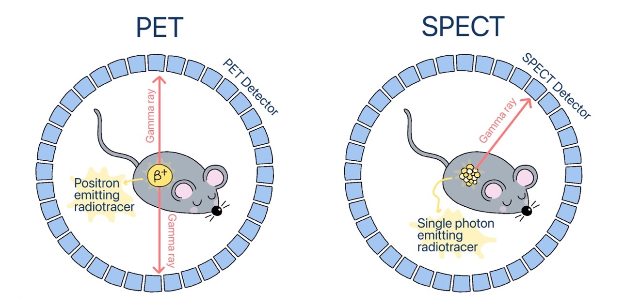
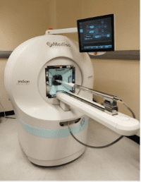
The nuclear medicine imaging modalities, Positron Emission Tomography (PET) and Single Photon Emission Computed Tomography (SPECT), utilize radioactive tracers to visualize and measure biological processes at molecular and cellular levels.
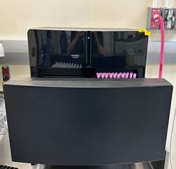
Hidex Gamma counter is ideal for nuclear medicine and PET applications, with precise onboard balance, samples can automatically be weighed for preclinical biodistribution studies. It can count up to 230 samples that are 13mm in diameter and 72 samples at 28mm in diameter. It is a compact system with lead shielding to minimize interference as well as detects energies from 15-2000KeV.
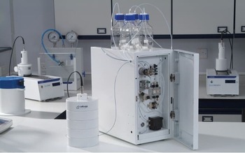
This compact HPLC system not only has a UV-Vis detector, but also a NaI scintillation detector for characterizing and quantifying radioligands. It is sensitive enough to read low amounts of radioactivity over a wide range of energies and allows the delivery of up to 4 solvents allowing for maximum flexibility. Using the Laura software, the researcher can create and edit methods, set up sample runs, and view data collection in real time.
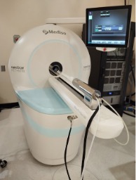
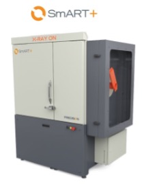
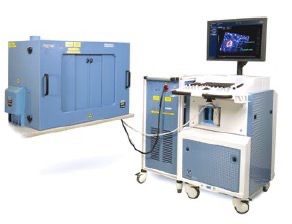
STTARR has installed the new SmART+ system, the small animal irradiator at PMCRT in Nov. 2022.
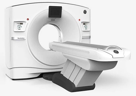
CT (Computed Tomography) combines a series of x-ray images taken from different angles around the subject or animal. Taking advantage of the differences in the x-ray attenuation properties of the various tissues and organs, computer processing creates cross-sectional images (slices) of the internal structures.
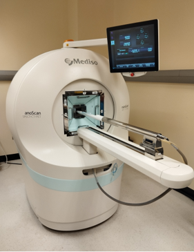
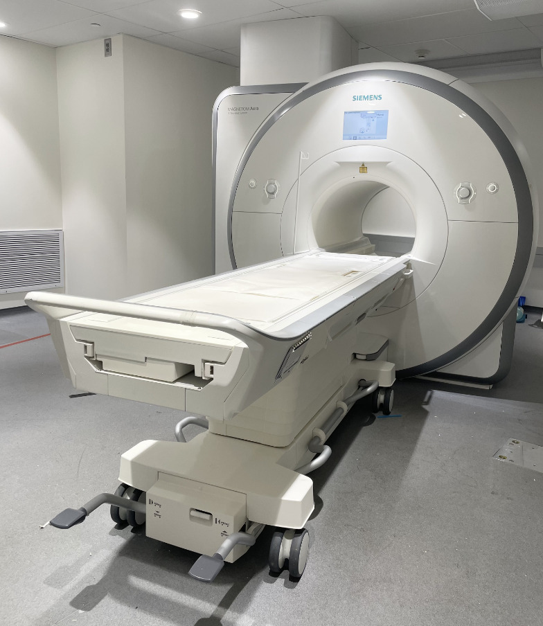
A MRI (Magnetic Resonance Imaging) scan uses the magnetization from water protons to generate images in which brightness can reflect not only the density of water protons in tissue but also the interaction of water protons with their local microenvironment. These images provide an unparalleled soft tissue contrast, enabling both anatomic and functional imaging for monitoring tumor growth and response to therapies.
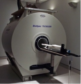
** The Bruker 7T MRI is now upgraded as of Spring 2023, to the NEO architecture running ParaVision 360, which will provide safer operation, higher throughput imaging, longer hardware lifetimes, and best-in-class fast imaging.
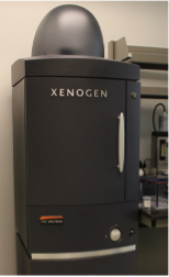
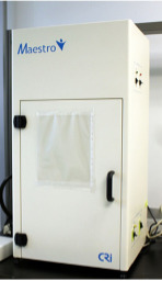
Fluorescence and Bioluminesence imaging techniques.
Real-time images are generated by applying high-frequency sound and then detecting the returning sound waves (echoes) caused by differences in acoustic impedence of the various tissue interfaces within the subject of interest. In addition to 2D B-mode images, anatomical 3D images of a small region can also be obtained. If the ultrasound beam is directed into a blood vessel, the blood flow gradient can be measured by a technique known as the Doppler effect. Visualization of blood flow can be further enhanced by the use of acoustic contrast agents such as microbubbles.
The photoacoustic (PA) effect refers to the generation of acoustic waves from an object being illuminated by pulsed or modulated electromagnetic (EM) radiation, including optical waves. The fundamental principle of the PA effect is based on the thermal expansion resulting from the absorption of EM radiation. The thermal expansion increases the acoustic pressure in the medium. Pulsing or modulating the EM radiation generates an acoustic wave which can be detected using an ultrasound transducer.
STTARR provides pre-clinical image analysis software through a variety of workstations that you can use autonomously or with STTARR staff assistance.
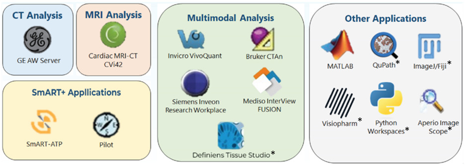
Note: Software marked with an asterisk (*) do not have staff support available. For software without the asterisk, our staff is available to assist you.